Loading...
Biomedical Imaging and Healthy Aging Laboratory
Who we are
We are a multidsciplinary reseach team of neuroscientists, clinicians, engineers, and mathematicians who study the brain with the shared goal to shed light on factors predicting conversion to neurodegenerative deseases.
More about us


What we do
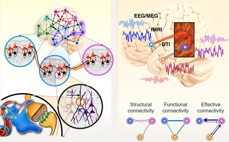
NeuroImaging & Signals
MRI • PET • EEG • ASL • Voice Analysis • Neuro-vascular coupling • Brain Mapping • Functional Connectivity • Structural Connectivity • Predictive Neuroimaging • Resting-state Networks
Brain Simulation
Whole-Brain Dynamic Models • BigBrain • Virtual Brain Activities • Neural Dynamics • Numerical Simulations • Artificial Intelligence • Deep Learning
Computational Models
Energy Metabolism • Tau Protein Accumulation • GABA • Glutamate • Neuro-Glia Vascular Model • Neurodegeneration
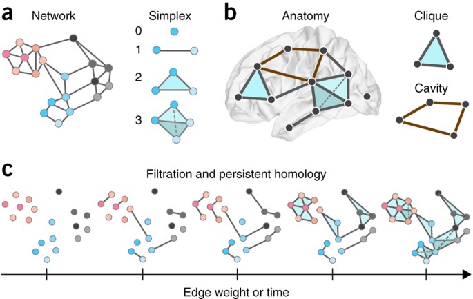
Mathematical Neuroscience
Algebraic Topology • Multilayer Networks • Complex Systems • Persistent Homology • Statistics • Information Theory • Spectral Theory • Network Neuroscience
Our team
Habib Benali, PhD
PI & Lab Director, Professor, Department of Electrical and Computer Engineering, Canada Research Chair in Biomedical Imaging and Healthy Aging, Member Applied AI Institute


Arsalan Rahimabadi, MSc
A brief introduction
Arsalan Rahimabadi is a Ph.D. candidate in the Electrical and Computer Engineering Department at Concordia University. His current research is focused on modeling tauopathy progression in the brain. He has expertise in dynamical systems analysis, pathological nonlinear systems, modeling, identification, control theories, and fault detection and isolation methods.
Educational background
He received his M.Sc. degree in Electrical Engineering – Control as the first-ranked student (out of 26) from K. N. Toosi Univ. of Tech. which is the industrial control center of excellence (ICCE) of Iran, and his B.Sc. degree in Electrical Engineering – Control as the first-ranked student (out of 114) of my faculty from Shahrood Univ. of Tech., Iran.
Professional background
Since Sep. 2019, he has been a researcher at PERFORM Centre (Montreal). From Jun. 2017 to Sep. 2019, he was the director of dynamical systems analysis and control team of Advanced Robotics and Automated Systems (ARAS) Research Group, Tehran, Iran. From Mar. 2013 to Apr. 2017, he was a researcher at ARAS Research Group.
Research
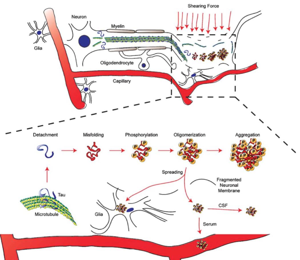
According to World Alzheimer Report, dementia has affected the lives of over 50 million people around the world, and this figure can rise threefold by 2050. Currently, dementia imposes a cost of trillion dollars annually, and if no cure is found, this figure can be doubled by 2030. The most prevailing form of dementia is Alzheimer's disease (AD). AD belongs to a group of neurodegenerative diseases known as tauopathies characterized by tau protein aggregation. Thus, we aim to address one of the crucial open problems concerning dementia, which is modeling tauopathy progression in the brain.
First discovered in 1975, tau is a microtubule-associated protein (MAP) in the neuron, which many researchers have extensively studied its function to stabilize microtubules and encourage axonal prolongation. This protein is natively unfolded, and in physiological conditions its tendency for aggregation is low. However, there are modifications, such as phosphorylation, which may enable tau proteins to make aggregates. The mechanisms and pathways by which tau protein forms aggregates in tauopathies are not sufficiently comprehended. Hence, our first step is to model the aggregation process of tau. Furthermore, since it has been experimentally proved that these aggregates do not remain in a specific part of the brain and they can spread all over the brain by axonal transportation, we also need to consider the diffusion process in our models. To accomplish our goal, we will take advantage of dynamical systems and control theories to analyze the proposed models.
Journal publication
- Rahimabadi, A. and Benali H. (2023). Extended fractional-polynomial generalizations of diffusion and Fisher-KPP equations on directed networks: Modeling neurodegenerative progression. bioRxiv
- Rahimabadi, A. and Benali H. (2022). Comment on the article “Spatially-extended nucleation-aggregation-fragmentation models for the dynamics of prion-like neurodegenerative protein-spreading in the brain and its connectome 486 (2020) 110102”. Journal of theoretical biology
- Rahimabadi, A., Taghirad H. D. (2017) “Comment on: “Centers of quasi-homogeneous polynomial planar systems,” [Nonlinear Anal.RWA 03 (6006) 409],” Nonlinear Analysis: Real World Applications
- Rahimabadi, A., Taghirad H. D. (2015) “Corner stability in nonlinear autonomous systems,” Nonlinear Dynamics
Conferences
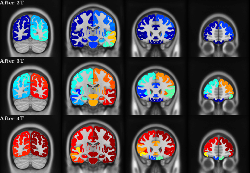
- Rahimabadi, A., Soucy JP., and Benali H. (2023). A computational model to study the combined effect of neuronal connectivity loss and tauopathy progression. HAI Conference
- Rahimabadi, A., Soucy JP., and Benali H. (2021). A comprehensive computational model of tauopathy progression using PET imaging. OHBM Conference
- Rahimabadi, A., Soucy JP., and Benali H. (2020). Effect of initial concentrations on a computational model of tau aggregation in tauopathies. OHBM Conference
Contact details
Email address: arsalan.rahimabadi@concordia.ca